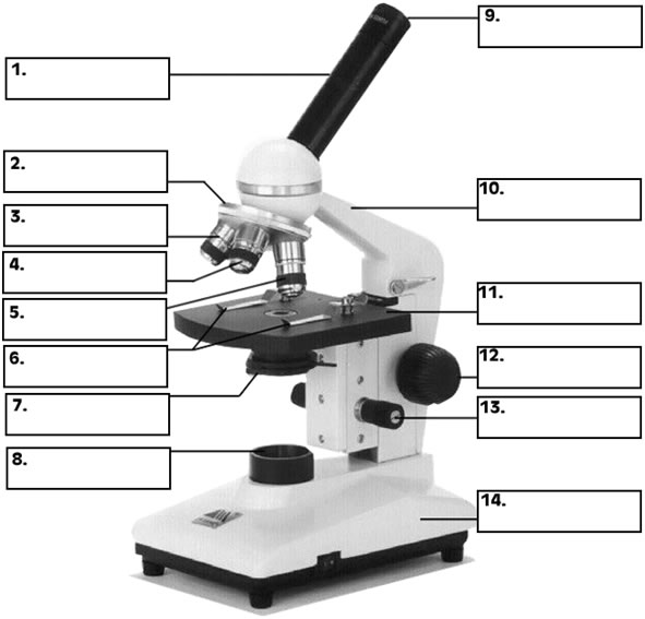
Fig.1.1: Parts of Compound Microscope.
|
Back Introduction
|
Home Cover Page
|
Top Lesson 1
|
Next Lesson 2
|
Lesson One: Microscope
Compound light microscope, iodine dye, dropper pipette, silk threads, microscope slides cover slips, newspaper, scissors, and onion.
Paramecium (unicellular organism) and bacteria (prokaryotic organisms).
Demonstration, short lecture and group work
The teacher may introduce the lesson by posing focus questions as like:-
The teacher may expect responses from students that may elaborate the concept of microscopy.
The basic concept to be discussed here is the role of microscope in viewing tiny organisms that could not be viewed with naked eyes. The microscope's invention and improvement in the seventeenth century led to the discovery and study of cells. In 1665, Robert Hooke, after studying a cork cell under light microscope, introduced the term cell. A microscope enables biologists to investigate living things and objects that are too small to be seen with the naked eye. The microscope is able to magnify these tiny specimens by means of lenses located in the eyepiece and objectives. The microscope is also capable of revealing fine details. This ability to reveal fine entity is known as resolving power.
Specimens that are viewed under the microscope are mounted on one of two types of glass slides. Prepared slides are slides that are for permanent use. Mount slides are for temporary use. Most of the slides that we use in biology will be wet mounts.
Demonstration of objects under a compound light microscope is required. Charts and diagrams that clarify those objects to be observed under the microscope should be posted in the corner of the laboratory room. This enables the students to identify the parts and compare their observations with actual diagram or picture of the objects or specimens.
The teacher is expected to demonstrate the cells under a microscope so that students can get the opportunity to observe them.
The students can begin working with familiar objects (e.g. a printed letter such as “e” or “f” to illustrate inversion properties of the microscope), and work up to easily viewed objects (e.g. permanent slides selected by the teacher or teacher prepared slides), and then to a variety of objects they select and prepare by themselves. A simple and familiar cell for microscopic observation might be onion epidermal cell. Therefore, it is highly recommended to observe onion epidermal cells using compound microscope.
The teacher should guide students how they can properly use a microscope. Therefore, in this investigation, the students should get the opportunity to:
Adjust the lower objective lens into viewing position.
NOTE. Always locate an object first with lower magnification even if a higher magnification is desired.
CAUTION:Never use the coarse adjustment wheel for focusing with high power. Damage to the lens and slide may result if the coarse adjustment wheel is used.
Students should be able to practice wet-mount and staining techniques, focusing techniques, and effective presentation of their observations to others. Diagrams of the specimens viewed should include field of view and magnification, should accurately show what they have seen.
Students should view a variety of plant and animal cells under the microscope, and use diagrams, models, or charts to illustrate and distinguish the structures of plant and animal cells.
Its position on the stage?
In what position does the letter move as seen through the microscope?
In what direction does the letter move as seen through the microscope?
In what direction does the letter move as seen through the microscope?
The following basic points should be noted.
Two important concepts in microscopy are magnificationand resolving power.
By the early 1900s, optics in light microscopes was good enough to achieve the best resolution, so improvements since then have focused on improving contrast. In the 1950s, researchers began to use the electron microscopewhich far surpassed the resolving power of the light microscope.
Resolving power is inversely related to wavelength. Modern electron microscopes have a practical resolving power of about 2 nm. Enhanced resolution and magnification allowed researchers to clearly identify sub cellular organelles and to study cell ultra structure using electron microscopes.
The teacher is supposed to summarize the lesson.
The students can match the following listed terms with their appropriate part of a microscope indicated with numbers and describe the use of each part.
Eyepiece or ocular, Body tube , Fine adjustment knob , Nosepiece, High power objective lens , Low power objective, Diaphragm , Mirror , Base , Coarse adjustment, Arm , Stage clips, Inclination joint, Middle objective.
Fig.1.1: Parts of Compound Microscope.