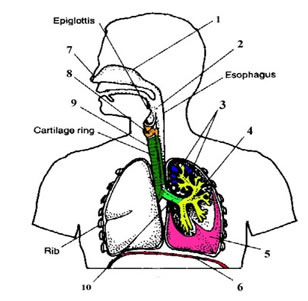
Fig. 8.1 The Human Breathing System
|
Back Lesson 7
|
Home Cover Page
|
Top Lesson 8
|
Next Lesson 9
|
Lesson Eight: The Human Breathing System
Suggested teaching Methodology
Lecture, Group work, Collaborative learning, Practical demonstration
The teacher may use different ways of introducing the lesson. For example, after posting the unlabelled diagram of the human breathing system, he/she can assess the priour knowledge of students by asking the following questions.
The above questions help to engage students and promote active participation. The teacher may expect possible answers as follows.
Fig. 8.1 The Human Breathing System
The breathing system comprises nostrils, trachea, bronchi, bronchioles and alveoli (alveolus). Air enters through nostrils or openings of the nasal passges and reaches trachea an then to bronchi which inturn branch into bronchioles, and finally air reaches alveoli ( air sacs).
Lecture method
The teacher is expected to explain how air from the atmosphere reaches the alveoli. The specific functions of nostrils, epiglottis, larnyx, trachea, alveoli etc should be described here.
Air from the atmosphere enters through the nostrils and is then filtered by hairs, warmed and Humidified, and sampled for odors as it flows through the nasal cavity. The nasal cavity leads to the phalarynx, an intersection where the paths for air and food cross. When food is swallowed, the larynx mu moves upward and tips the epiglottis over the glottis. The rest of the time, the glottis is open, and air enters the upper part of the respiratory tract. The wall of the larynx is reinforced by cartilage. From the larynx, air passes into the trachea, or windpipe, whose shape is maintained by rings of cartilage. The trachea forks into two bronchi, one leading into each lung. Within the lungs, each bronchus branches repeatedly into finer and finer tubes, called bronchioles. At their tips, the thinnest bronchioles dead-end as a cluster of air sacs called alveoli. The epithelium lining the major branches of the respiratory tree is covered by cilia and a thin film of mucus. The mucus traps dust, pollen, and other particulate contaminants, and the beating cilia move the mucus upward to the pharynx, where it is swallowed.
What specific functions are performed by the nasal cavity?
As air passes through the nasal passages, the following major activities are performed.
The muscular pharynx (throat) carries air into the respiratory tract and foods and liquids into the digestive system. Thus, the pharynx serves as a common passage for food and air.
The larynx (voice box) is located between the pharynx and the trachea. It has a framework of cartilage that protrudes in the front of the neck. At the upper end of the larynx are the vocal cords, which serve in the production of sound. They are set into vibration by the flow of air from the lungs. The space between the two vocal cords is called the glottis, and the little leaf-shaped cartilage that covers the larynx during swallowing of food is called the epiglottis. The epiglottis prevents food from entering into trachea.
How is it possible for epiglottis to prevent food from entering into trachea?
As the larynx moves upward and forward during swallowing, the epiglottis moves downward, covering the opening into the larynx. You can feel the larynx move upward toward the epiglottis during this process by placing the flat ends of your fingers on your larynx as you swallow.
Where does gas exchange take place?
GasGas exchange occurs across the thin epithelium of the lung’s millions of alveoli. These have a total surface area of about 100 m2 in humans, sufficient to carry out gas exchange for the whole body.
Oxygen in the air entering the alveoli dissolves in the moist film and rapidly diffuses across the epithelium into a web of capillaries that surrounds each alveolus. Carbon dioxide diffuses in the opposite direction from blood to alveoli.
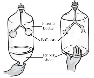
Fig. 8.2: Model to show the Mechanism of Breathing
The teacher can engage the students by posing different stimulating questions like:
You are aware that the air we inhale or exhale is a mixture of gases. What do we exhale? Do we exhale only carbon dioxide or a mixture of gases along with it? You must have also observed that if you exhale on a mirror, a film of moisture appears on its surface. Where do these droplets come from? To understand the expansion of the lungs, pull the rubber sheet from the base downwards and watch the balloons. Next, push the rubber/plastic sheet up and observe the balloons. Did you see any changes in the balloons?
What do the balloons in this model represent? What does the rubber sheet represent? Now, the students should be able to explain the mechanism of breathing.
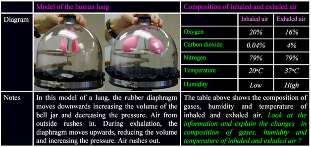
Fig. 8.3: Model of Human Lung
Ask students to explain the mechanism as to how breathing takes place (how inhalation and exhalation are effected). This topic should be supported by simple practical activity/demonstration. Let the students be grouped into smaller groups and explain the mechanism of breathing. For practical demonstration, you can follow the steps found in your textbook. Lung volume increases as a result of the contraction of the rib muscles and diaphragm, a sheet of skeletal muscle that forms the bottom wall of the chest cavity. Conversely, the volume of the chest cavity decreases during the relaxation of the rib muscles and diaphragm.
What happens during inhalation or breathing in?
During Inhalation,
What happens during Exhalation or breathing out?
During exhalation,
During vigorous exercise, other muscles of the neck, back, and chest further increase ventilation volume by raising the rib cage even more.
| Lung volumes | Definition | Average amount of air in cc |
|---|---|---|
| Tidal volume | The amount of air moved into or out of the lungs during normal breathing | 500 cc |
| Vital capacity | The volume of air that can be expelled from the lungs by maximum exhalation following maximum inhalation | 4800 cc |
| Residual volume | The volume of air that remains in the lungs after maximum exhalation | 1200 cc |
| Total lung capacity | The total volume air that can be contained in the lungs after maximum inhalation | 6000 cc |
Table: 8.3. Breathing Volumes
The teacher is required to ask features of alveoli to allow efficient gaseous exchange.
The features of alveoli for which allow them for efficient gas exchange include:
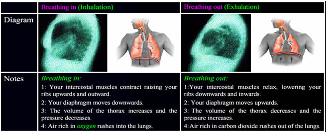
Fig . 8.4: Model Demonstrating Inhalation and Exhalation
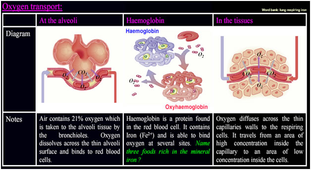
Fig. 8.5. Diffusion and Transport of oxygen
The students are also able to explain the features of blood capillaries that allow them for efficient gas exchange which include: